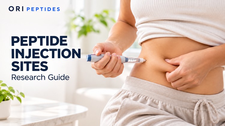
Peptide injection sites are the body regions named in study protocols where a peptide solution is placed under the skin. Protocols name the site, record it, and track what happens there, because published research shows that the site can affect tissue depth, absorption, and local reactions. This guide explains what the research literature records and why. It describes published studies only and is not advice or instruction for any person.
Many online pages about this topic are written as how-to guides for people, and they come from telehealth companies, pharmacy brands, wellness publishers, and vendors. This guide does not follow that format. It sticks to what published papers measured and leaves out any claim that could not be traced to one.
What Are Peptide Injection Sites in Research?
A peptide is a short chain of amino acids. Many research peptides arrive as a freeze-dried powder and are mixed with liquid before a study uses them. When a study delivers the solution under the skin, the route is called subcutaneous. Subcutaneous means into the fat layer that sits between the skin and the muscle underneath.
The regions that published studies name for this route are the belly area (abdomen), the thigh, the upper arm, and for some drugs the buttocks. A protocol that names one of these regions is describing where the solution was delivered in that study, so that someone else can compare or repeat the work.
One point matters before anything else. Most of the published data on injection sites come from daily-use drugs such as insulin, and from a few other drugs made of peptides or proteins. The papers reviewed for this guide did not include a study that tested injection sites for the research compounds sold for laboratory use. What follows is the closest published evidence, and it should not be assumed to apply to any other compound.
What a Protocol Lists as Supplies
Before a study starts, its protocol lists the materials, so that the work can be repeated and checked. For a peptide delivered under the skin, the technique literature and the concentration calculator page name the same kinds of items. The list below describes what protocols name. It is not a shopping list for any person.
| Item | What it is | Why a protocol lists it |
|---|---|---|
| Freeze-dried peptide vial | The compound as a sealed dry powder | Strength and lot number tie results to a batch |
| Certificate of Analysis | Lab paperwork showing identity and purity | Confirms what the vial holds |
| Bacteriostatic water | Sterile water with a small amount of preservative | Used to mix a vial that will be sampled more than once |
| Syringes | Marked on a U-100 scale, in 0.3, 0.5, or 1 mL sizes | Barrel size affects how precisely a volume can be read |
| Needles | Listed by length | Length affects how deep the solution goes |
| Alcohol swabs and gloves | Cleaning and hand protection | Cleanliness steps are recorded |
| Labels | For the date mixed and the concentration | Lets the mixed solution be traced |
| Sharps container | A puncture-proof container for used needles | Disposal is recorded |
How Protocols Describe Reconstitution
Reconstitution means adding liquid to a dry powder so it becomes a solution that can be measured. The calculator page and the peptide reconstitution guide describe the usual laboratory method in the same order. Both stoppers are swabbed and left to dry. The water is drawn and added slowly down the inside wall of the vial, because a fast stream onto the powder causes foaming. The vial is swirled gently and never shaken, since shaking can damage peptides, and the liquid should end up clear. The vial is then labeled with the date and the concentration, and stored in a refrigerator.
How long a mixed vial stays usable depends on the compound, and the product’s own paperwork sets that window, so no number is given here. The bacteriostatic water guide explains why preserved water is the usual choice. A protocol record for this step lists the water volume, the date and time of mixing, the resulting concentration, and the storage conditions.
See also: Peptide Reconstitution Ratios & How to Calculate Them
How Protocols Work Out the Amount
Protocols that list an amount also show the math behind it, so that anyone can check it. The calculator page carries three steps through to a syringe reading.
- Concentration equals the milligrams in the vial divided by the milliliters of water added.
- Draw volume equals the amount in milligrams divided by the concentration.
- Units on a U-100 syringe equal the draw volume in milliliters multiplied by 100.
A U-100 syringe has 100 units to every milliliter, so 10 units is 0.1 mL on any barrel size. The calculator page shows the worked example below only to illustrate the arithmetic, using a 5 mg vial mixed with 2 mL of water and an amount of 1 mg.
| Step | Calculation | Result |
|---|---|---|
| Concentration | 5 mg ÷ 2 mL | 2.5 mg per mL |
| Draw volume | 1 mg ÷ 2.5 mg per mL | 0.40 mL |
| Syringe reading | 0.40 mL × 100 | 40 units |
Two habits from the calculator page keep the math clean. Concentration is set once, when the water goes in, so changing the water volume means working it out again from the start. An amount listed in micrograms is converted to milligrams by dividing by 1,000, once, before the first formula. This section explains arithmetic and record keeping. It does not set an amount for any compound or any person.
How Protocols Record the Draw
The draw is the volume of liquid pulled into the syringe, and protocols record it in milliliters and often in units as well. Several details affect how accurate that reading is, and the calculator page lists them.
Syringe size. All three common sizes use the same 100-units-per-milliliter scale, but a smaller barrel spreads the scale over a shorter length. The page notes that the smallest barrel that comfortably holds the volume gives the most precise reading, so records name the size used.
Reading the barrel. The page advises reading the volume at eye level and not from an angle, and clearing air bubbles, since both are common sources of small errors.
Dead volume. This is the small amount of liquid left in the hub and needle after the plunger is fully pressed. It is part of why manufacturers add a little extra powder to a vial, and the labeled strength is what the calculations assume.
These are measuring habits, and the calculator page describes them for vial math. They are not directions for any person.
Why Site Selection Appears in Protocols
Three findings in the literature explain why protocols name the site.
Tissue depth differs by site. A 2010 study measured skin and fat thickness with ultrasound at four sites in 388 US adults. Skin was about 2 mm thick everywhere. The fat layer was thicker in some places than others, and it was also thicker in women, by about 5 mm on average, and in people with a higher body mass index. The study was run by authors who worked for a medical device company, and it covered only US adults with diabetes, so the numbers are a guide and not a rule.
Absorption can differ by site, though not always. A 2003 study looked at a peptide drug given under the skin twice a day. Twelve adults used three sites in turn, each for about a week. Using the abdomen as the starting point, the amount of drug reaching the blood was 101% for the thigh and 117% for the upper arm. The authors saw the three sites as comparable for absorption. This is one drug in 12 people, so it does not show how another peptide would behave.
Local reactions can differ by site. The same 2003 study found that skin reactions were common but mild, and that how often they happened and what they looked like varied with the site. This is one reason studies track the site next to every reaction they log.
| Site named in the studies | Average fat layer thickness, 2010 adult study | Drug absorbed compared with the abdomen, 2003 study |
|---|---|---|
| Abdomen | 13.9 mm | 100% (the starting point) |
| Thigh | 10.4 mm | 101% |
| Upper arm | 10.8 mm | 117% |
| Buttocks | 15.4 mm | Not tested |
The two studies measured different things in different groups of people. The table puts them side by side only to show why a protocol that says “subcutaneous” without naming a site leaves out useful information.
See also: Semaglutide Dosage and Reconstitution for Research
What Subcutaneous Study Routes Describe
When a protocol describes a subcutaneous route, the published papers show a set of details that are written down, so that the work can be repeated.
The named site and side. Protocols name the region and note left or right, because the next record refers back to it.
Needle length. The 2010 adult study found that needles 8 mm or longer, placed straight in, could frequently reach muscle in the limbs of men and in people with a lower body mass index. Needles of 4 to 5 mm stayed in the fat layer in almost all adults in that study. This is a finding about tissue depth, and it is the reason needle length is recorded and not only the site.
Skin handling. Expert workshops on injection technique, held from 1997 on, have written recommendations that cover skin folds and shorter needles. A workshop summary from 2010 describes the history of that work. These are written for a clinical setting, and they show how much detail the technique literature records.
The solution itself. Concentration, amount, and syringe size are recorded alongside the site. The sections above explain how those numbers are worked out.
None of this tells any person how to give an injection. It lists what published work chose to write down.
Rotation in Repeated Dosing Studies
Rotation means changing the site from one entry to the next. It appears in the literature because of a skin and fat change called lipohypertrophy, which is a lump of thickened fat that forms at a site that is used again and again. Here is what the papers show.
- A 2021 study of 316 insulin users. Ultrasound found hidden lipohypertrophy in 19.9% of them, even though it could not be felt or seen by checking the skin. The study linked it with incorrect site rotation, with reusing needles more than four times, and with a higher body mass index. People with the hidden lumps also had worse blood sugar control, which is why they matter in an insulin study.
- A 2012 case report of two women. Both had thickened fat at the belly site used for a protein drug. The thickened fat shrank after the site was moved to the thighs, and it came back at the new site. The authors suggested that rotation might help prevent it, and noted that the strength of the evidence is weak.
- A 2016 prevention paper. It lists site rotation, patient education, and avoiding needle reuse among the ways to reduce lipohypertrophy, and it is a short expert view and not a trial.
The limit is that these results involve drugs used daily for months or years. They show why repeated-dosing protocols write down the site of every entry. They do not show how any research compound behaves, and none of the papers tested one.
Documentation Researchers Keep After Each Entry
Good records let another person see what happened, in what order, and where. A review of 158 papers on injection site reactions in trials of biologic drugs given under the skin found that the most common reactions were redness (42.8%), pain (12.4%), and itching (5.7%). How often reactions were reported ranged from under 1% to 15.5%, depending on the drug. The authors called for better reporting of these events, which is an argument for careful notes in any study.
The table lists the fields that protocols and the literature commonly track, and why each one is useful.
| Record field | Why it is recorded |
|---|---|
| Date and time | Lets entries be put in order |
| Compound and lot number | Ties the result to a specific batch |
| Vial strength and water volume | Sets the concentration of the mixed solution |
| Amount and draw volume | Shows exactly what was delivered |
| Syringe size and needle length | Affects reading accuracy and tissue depth |
| Named site and side | Allows comparison and rotation tracking |
| Previous site | Shows the order of sites used |
| Skin notes | Logs redness, swelling, itching, pain, or lumps |
Notes on the skin, such as lumps, are recorded at every entry, because the studies above found that lumps can be missed unless someone looks for them. The record does not stop at the entry itself. Protocols also note where the used needle went, since the 2021 study linked reusing a needle more than four times with hidden lumps, and they note that the mixed vial went back to storage with its label updated.
See also: BPC-157 Dosage and Reconstitution Research Guide
Frequently Asked Questions
What are peptide injection sites?
They are the body regions named in study protocols where a peptide solution is placed in the fat layer under the skin. The regions named in published studies include the abdomen, thigh, upper arm, and for some drugs the buttocks.
Why do protocols name the site?
Published studies found that tissue depth, absorption, and local reactions can differ by site. Naming the site lets others compare or repeat the work.
Does the site change how much is absorbed?
In one 2003 study of a peptide drug in 12 adults, absorption was 101% for the thigh and 117% for the upper arm compared with the abdomen. That result is for one drug and may not apply to others.
Why does rotation appear in the research?
Repeated use of one site can cause lipohypertrophy, a lump of thickened fat. A 2021 study found hidden lumps in 19.9% of 316 insulin users and linked them with incorrect rotation and needle reuse.
What do researchers record about each entry?
Protocols commonly list the date, compound and lot, concentration, amount, syringe and needle details, named site and side, previous site, and any skin notes.
Is there research on injection sites for research compounds?
The papers reviewed for this guide did not include a study that tested injection sites for research-grade peptides. The available evidence comes from daily-use drugs.
What supplies do protocols list?
They list the vial and its Certificate of Analysis, bacteriostatic water, syringes and needles by size and length, swabs, labels, and a sharps container. Each item is listed so the work can be checked and repeated.
How do protocols describe reconstitution?
They describe adding preserved water slowly down the vial wall, swirling gently without shaking, checking that the liquid is clear, and labeling the vial with the date and concentration before refrigerating it.
How are syringe units worked out?
Concentration is the milligrams in the vial divided by the milliliters of water added. Draw volume is the amount divided by the concentration, and units on a U-100 syringe are the draw volume times 100. The concentration calculator does this for you.
Key Takeaways
- What they are. Peptide injection sites are the named body regions where a study delivers a solution under the skin.
- Why they matter. Fat thickness, absorption, and local reactions can differ by site, based on studies of daily-use drugs.
- Supplies and mixing. Protocols list each item and record the water volume, mixing date, concentration, and storage.
- The math. Concentration, draw volume, and U-100 units follow three steps that the concentration calculator carries out.
- Needle length. Protocols record it because it affects how deep the solution goes.
- Rotation. It appears in the literature because repeated use of one site can cause lipohypertrophy, and hidden lumps were found in 19.9% of one group.
- Documentation. Records list the date, compound, lot, concentration, amount, syringe, needle, site, previous site, and skin notes.
- The limit. No paper reviewed tested injection sites for research compounds, so none of this sets a procedure for any person.
Research Use Disclaimer
This content is provided for Research Use Only (RUO). The compounds discussed are not drugs, dietary supplements, foods, or cosmetics, and are not intended for human or animal consumption, diagnostic use, or therapeutic use of any kind. They have not been evaluated by the FDA for safety or efficacy in humans. Any research use must be conducted by qualified professionals in a controlled laboratory environment, in accordance with all applicable institutional, local, and federal regulations.
Sources
- Lalezari JP, Patel IH, Zhang X, et al. Influence of subcutaneous injection site on the steady-state pharmacokinetics of enfuvirtide (T-20) in HIV-1-infected patients. J Clin Virol. 2003.
- Gibney MA, Arce CH, Byron KJ, Hirsch LJ. Skin and subcutaneous adipose layer thickness in adults with diabetes at sites used for insulin injections, implications for needle length recommendations. Curr Med Res Opin. 2010.
- Frid A, Hirsch L, Gaspar R, et al. The Third Injection Technique Workshop in Athens (TITAN). Diabetes Metab. 2010.
- Luo D, Shi Y, Zhu M, et al. Subclinical lipohypertrophy, easily ignored complications of insulin therapy. J Diabetes Complications. 2021.
- Rochira V, Zirilli L, Diazzi C, Romano S, Carani C. Clinical and radiological evidence of the recurrence of reversible pegvisomant-related lipohypertrophy at the new site of injection in two women with acromegaly, a case series. J Med Case Rep. 2012.
- Kalra S, Kumar A, Gupta Y. Prevention of lipohypertrophy. J Pak Med Assoc. 2016.
- Kim PJ, Lansang RP, Vender R. A systematic review and meta-analysis of injection site reactions in randomized-controlled trials of biologic injections. J Cutan Med Surg. 2023.
Explore the research peptide catalog for laboratory compounds, and use the concentration calculator for concentration math. For related reading, see the peptide reconstitution guide and the bacteriostatic water guide.